


1
2
3
4
5
6
7
8
9
10
11
12
13
14
15
16
17
18
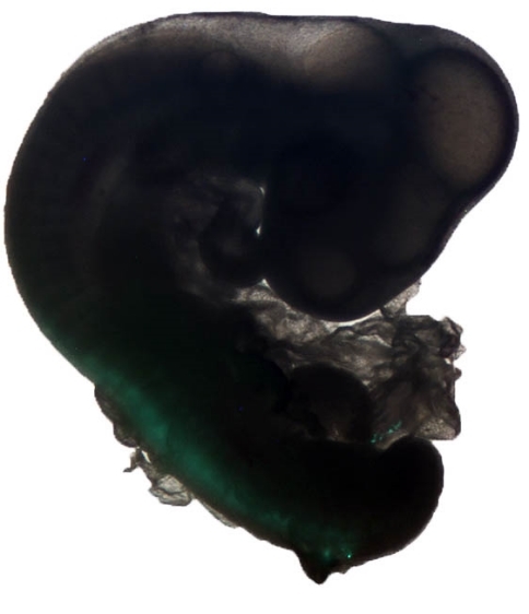
HH21 chick expressing GFP
Reconstructed μ-CT Scan of Gecko Ver
micro-CT scan of a gecko vertebra showing the autotomy plane. The demineralized gap in the centre of the vertebra breaks during autotomy to facilitate tail loss.
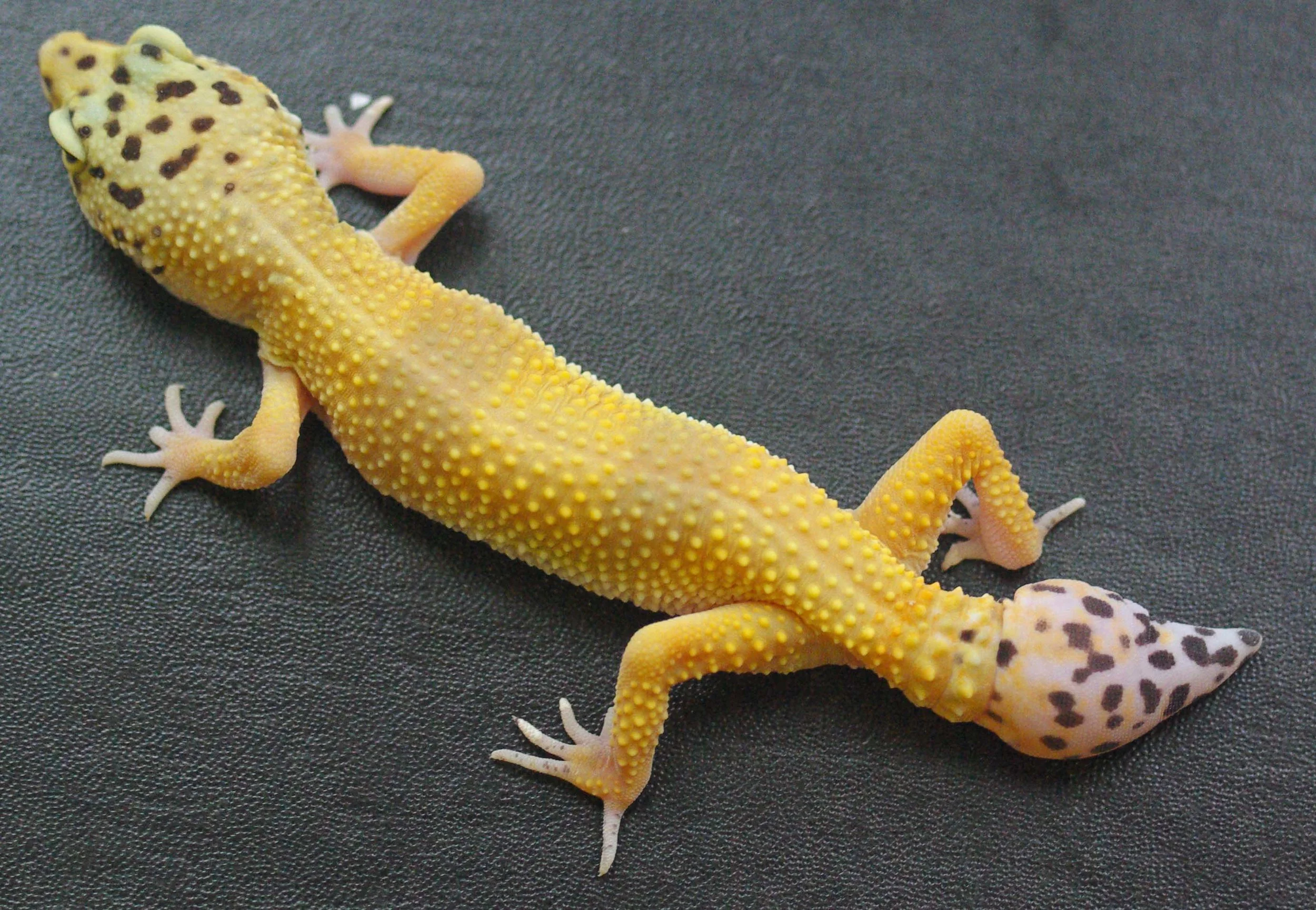
Regenerate tails
From right to left: Original tail, a stage III regenerate tail, a stage VI regenerate tail, a fully regenerated (stage VII) tail, a very fat fully regenerate tail. Geckos store much of their body fat in their tails, and regenerate tails can become particularly fat.
Wound healing following autotomy
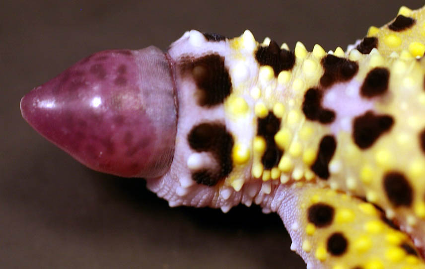
Regenerating tail
The pink cone protruding from the end of the tail stump is the blastema, covered blastema wound epidermis. This will form the regenerate tail.
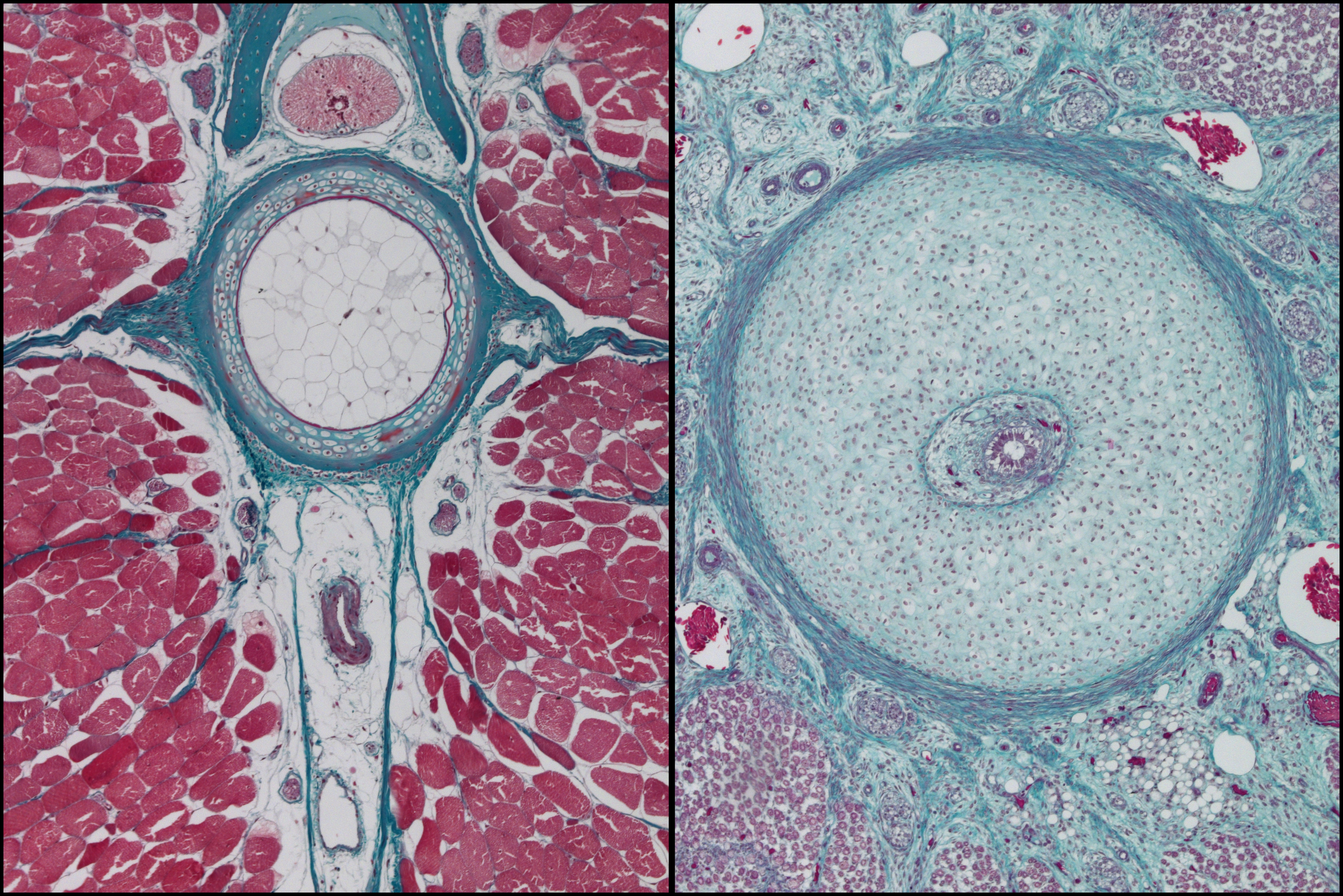
Original and Regenerated Gecko Tails
Masson's trichrome image of a transverse section through original (left) and regenerate (right) gecko tails. Muscle is stained red. Skeletal and connective tissue is blue-green.
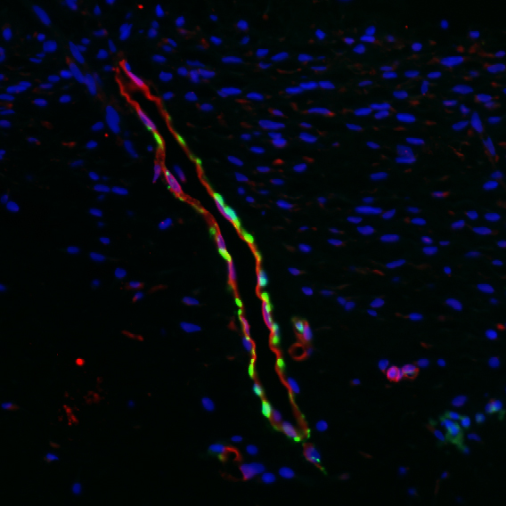
Small vessel supported by pericytes
Immunofluorescence image highlighting a small vessel. Blue is DAPI, showing nuclei of all cells. Red localizes von Willebrand factor to show the endothelial cells (inner lining) of the blood vessel. Green localizes α-smooth muscle actin, which is a marker vascular support cells, including the pericytes seen here.
Vein and Caudal Artery
Immunoflourescence image showing a vein (left, note the valve) and the caudal artery (right) of a gecko. Blue (faint) is DAPI staining for cell nuclei. Green localizes α-smoothe muscle actin, a marker for vascular smooth muscle cells, and red localizes von Willebrand factor, a marker of endothelium (the inner lining of blood vessels).
Blood vessels surrounding regenerate ependymal tube
Immunofluorescence image of blood vessels surrounding the regenerated ependymal tube. DAPI (blue) was used to stain nuclei. The ring of blue nuclei in the centre is the regenerate ependymal tube (spinal cord). Red localizes von Willebrand factor, a marker for the endothelial cells that line the inside of blood vessels. Green localizes α-smooth muscle actin, which marks vascular support cells (pericytes). In the topmost vessel, two green auto-fluorescent erythrocytes (red blood cells) are clearly visible. Unlike mammals, erythrocytes in reptiles (and birds) are nucleated.
Original tail histology
Transverse section of an original gecko tail (Chondrodactylus bibronii), stained with H&E